

The featured image and supporting patient illustrations are AI-generated and do not depict actual patients.
ASPC Patient Education · Updated 26 October 2026
At a glance
Heel pain may come from the plantar fascia, Achilles tendon, heel fat pad, bone or nerves. First-step pain under the heel is common in plantar fasciopathy, but the exact cause should be assessed before choosing exercises.
General patient information. Diagnosis and treatment should be individualized by a qualified healthcare professional.
Get help promptly: A sudden snap, inability to walk or a hot swollen foot needs urgent assessment.
In this guide
- What Is Heel Pain?
- Anatomy and Structures Involved
- How Common Is Plantar Heel Pain?
- Common Causes and Risk Factors
- Symptoms of Heel Pain
- Heel Pain: Pattern and Possible Cause
- Red Flags: When to Seek Medical Assessment
- How Is Heel Pain Assessed?
- Differential Diagnosis: Plantar Fasciopathy vs Achilles Tendinopathy
- Why Choosing the Right Treatment Matters
- Physiotherapy for Heel Pain
- Heel Pain Exercises
- Managing a Flare-Up
- Prevention and Self-Management
- Structural Diagnosis and Management (SDM) Perspective
- When to See a Physiotherapist or Doctor
- Final Takeaway
- Common questions
- References and clinical guidance
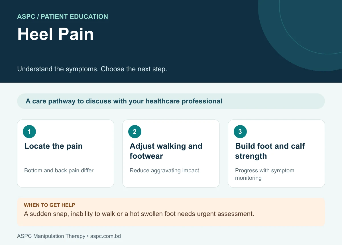
- Locate the pain: Bottom and back pain differ.
- Adjust walking and footwear: Reduce aggravating impact.
- Build foot and calf strength: Progress with symptom monitoring.
Heel pain is a common foot problem that can affect people of different ages and activity levels. It may limit walking, standing, work and sport. Plantar fasciopathy (often called plantar fasciitis) is a common cause of pain under the heel, but pain can also arise from the Achilles tendon, heel fat pad, bone, nerves or referred sources. Identifying the location, timing and behaviour of symptoms helps guide appropriate assessment and treatment. Most uncomplicated plantar heel pain can be managed with education, load adjustment and progressive rehabilitation (Koc et al., 2023).

The featured image and supporting patient illustrations are AI-generated and do not depict actual patients.
What Is Heel Pain?
Heel pain is discomfort in or around the heel bone (calcaneus). It can be felt on the underside, at the back, or along the inner or outer edge of the heel. Heel pain is a symptom rather than a diagnosis. The likely cause depends on the exact site, whether symptoms began suddenly or gradually, the amount of weight-bearing activity, and whether there are signs of nerve involvement or systemic illness.
Plantar heel pain is often associated with the plantar fascia, a strong band of connective tissue running from the heel bone towards the toes and helping support the foot arch. The term plantar fasciopathy is commonly used because persistent symptoms may involve degenerative and load-related changes as well as local irritation, rather than simple inflammation alone.
Anatomy and Structures Involved
The heel contains the calcaneus, a cushioning fat pad, the plantar fascia, the Achilles tendon insertion, nearby bursae, nerves and surrounding muscles and ligaments. These structures help absorb impact, transfer force and support walking and running.
Plantar fascia: supports the longitudinal arch and helps manage load during walking and running.
Calcaneal fat pad: cushions the heel during contact with the ground.
Achilles tendon: connects the calf muscles to the back of the heel and contributes to push-off.
Calcaneus: the heel bone can develop a stress injury after repetitive loading or fracture after trauma.
Nerves: compression or irritation can cause burning, tingling, numbness or shooting pain.
How Common Is Plantar Heel Pain?
Plantar fasciopathy is a frequent cause of plantar heel pain in adults and is particularly relevant to people whose work or recreation involves prolonged standing, walking or running. It may occur in physically active people as well as people with lower activity levels. Symptoms can persist or recur, so treatment should address the person's loading pattern, strength, mobility, footwear needs and functional goals rather than focusing only on the painful spot (Koc et al., 2023).
Common Causes and Risk Factors
Different conditions can produce similar heel symptoms. The following are important possibilities, but the list is not a substitute for a clinical examination.
Plantar fasciopathy: pain under the inner heel, often worst with the first steps after sleep or rest and sometimes easing after moving.
Achilles tendinopathy: pain and tenderness at the back of the heel or along the tendon, often aggravated by running, hills, jumping or repeated calf loading.
Heel fat-pad pain: deep, bruise-like pain under the centre of the heel, often worse on hard surfaces or with direct pressure.
Calcaneal stress fracture: progressive heel pain after a sudden increase in running, walking or impact activity; weight-bearing may become increasingly painful.
Nerve entrapment: burning, tingling, electric pain or altered sensation around the heel or sole.
Retrocalcaneal bursitis or Haglund-related irritation: pain, swelling or rubbing at the back of the heel, sometimes aggravated by shoe pressure.
Referred pain or systemic disease: symptoms may occasionally be linked to the back, inflammatory disease, infection or other medical conditions.
Potential contributors to plantar heel pain include a recent increase in walking or running, limited ankle dorsiflexion, reduced calf or foot-muscle capacity, prolonged weight-bearing, footwear that is poorly tolerated, and higher body mass in some non-athletic individuals. These factors influence risk but do not prove the cause in an individual (Koc et al., 2023).
Symptoms of Heel Pain
Symptoms vary according to the affected structure. Common features include:
Pain under, behind or along the sides of the heel.
Sharp pain with the first steps in the morning or after sitting, typical of plantar fasciopathy.
Pain that increases after prolonged standing, walking, running or jumping.
Tenderness when pressing the heel or the Achilles tendon.
Stiffness in the foot or calf, or difficulty walking normally.
Burning, tingling or numbness when a nerve is involved.
Swelling, bruising or sudden loss of weight-bearing ability in some injuries.
The pattern of pain provides useful clues, but it cannot confirm a diagnosis on its own. For example, first-step pain is common in plantar fasciopathy, whereas posterior heel pain during calf loading may suggest Achilles tendon involvement.
Heel Pain: Pattern and Possible Cause
| Pain location / pattern | Typical features | Possible causes |
|---|---|---|
| Bottom / inner heel | First-step pain after rest; tenderness near plantar fascia attachment | Plantar fasciopathy; sometimes fat-pad or nerve pain |
| Back of heel | Pain with calf raises, running or shoe pressure; Achilles tenderness | Achilles tendinopathy; bursitis; insertional irritation |
| Centre of heel | Bruised feeling, worse on hard surfaces or direct pressure | Heel fat-pad pain |
| Deep, progressive pain | Often follows increased impact; weight-bearing becomes painful | Calcaneal stress fracture |
| Burning or tingling | Pins and needles, numbness or shooting pain | Nerve irritation or entrapment |
Red Flags: When to Seek Medical Assessment
Seek urgent medical care if heel pain follows a major injury, the foot looks deformed, you cannot walk or bear weight, or you heard a snap with sudden severe pain. Prompt assessment is also important for a hot, red or markedly swollen heel, fever or other signs of infection. An open wound or ulcer around the heel—especially in a person with diabetes, poor circulation or reduced sensation—requires timely medical attention. Do not try to exercise through these warning signs (NHS guidance).
Arrange clinical review if pain is severe, worsening, repeatedly returning, associated with tingling or loss of sensation, or not improving after around two weeks of sensible self-care. People with diabetes should seek advice early because foot complications can be more serious (NHS guidance).
How Is Heel Pain Assessed?
A physiotherapist or clinician begins by asking about the exact location, onset, recent changes in walking or training, work demands, footwear, previous injuries and medical history. They may ask whether pain is worst with the first steps in the morning, after prolonged activity, during calf loading or when pressing the heel.
Observe walking, standing tolerance and foot/ankle movement.
Palpate the plantar fascia attachment, heel fat pad, Achilles tendon and other relevant structures.
Measure ankle dorsiflexion and assess calf flexibility, foot/ankle strength and heel-raise capacity.
Assess sensation and relevant neurological signs if burning, tingling or numbness is reported.
Use functional measures such as walking, single-leg balance or heel raises when safe and appropriate.
Imaging is not routinely required for a typical presentation of plantar fasciopathy. X-ray, ultrasound, MRI or other investigations may be considered when the presentation is atypical, a stress fracture or other pathology is suspected, symptoms persist despite appropriate care, or the result would change management. A heel spur on an X-ray does not by itself prove the source of pain.
Differential Diagnosis: Plantar Fasciopathy vs Achilles Tendinopathy
| Feature | Plantar fasciopathy | Achilles tendinopathy |
|---|---|---|
| Pain site | Bottom/inner side of heel | Back of heel or Achilles tendon |
| Typical aggravation | First steps after rest; prolonged weight-bearing | Running, hills, jumping or repeated calf loading |
| Tenderness | Near plantar fascia attachment | Along tendon or at tendon insertion |
| Rehabilitation emphasis | Plantar fascia and calf stretching, progressive foot/ankle loading | Progressive calf strengthening and load management |
Why Choosing the Right Treatment Matters
Heel pain is not one single condition. Treating every case as plantar fasciitis can delay appropriate care for a stress fracture, nerve problem, Achilles disorder or infection. A treatment plan should reflect the clinical findings and the person's daily demands. Long periods of complete rest may reduce capacity, while continuing high-impact activity despite worsening pain may overload the affected tissues. The aim is to adjust aggravating loads while maintaining safe activity and gradually rebuilding capacity.
Physiotherapy for Heel Pain
The 2023 clinical practice guideline supports a combined approach for plantar heel pain/plantar fasciitis, including education, plantar fascia-specific and calf stretching, appropriate manual therapy, taping for short-term relief and progressive foot/ankle strengthening. Orthoses may help when combined with other treatments, but should not be relied upon as the only short-term treatment. Night splints may be considered for people who consistently experience first-step pain in the morning (Koc et al., 2023).
Assessment and education: explain the likely diagnosis, recovery expectations and how to modify aggravating activities.
Load management: temporarily reduce running, jumping or long standing that substantially increases pain; build back gradually.
Stretching: use plantar fascia-specific and gastrocnemius/soleus stretching when indicated.
Strengthening: progressively train calf, foot and ankle muscles according to tolerance.
Manual therapy: joint or soft-tissue techniques may be used when relevant mobility restrictions are present, alongside exercise.
Taping or orthoses: taping may provide short-term symptom relief; shoe inserts may be considered as part of a combined plan.
Night splint: consider a 1–3 month programme for persistent first-step morning pain, guided by a clinician.
Monitoring: reassess pain, walking tolerance, heel-raise capacity and patient-specific goals; adjust the programme as function improves.
Heel Pain Exercises
Exercise selection should match the diagnosis. Do not use the same programme for every cause of heel pain. The following examples are commonly considered for plantar heel pain when fracture, acute tendon rupture and other contraindications have been excluded. A physiotherapist can modify the range, repetitions and progression.
1. Plantar Fascia-Specific Stretch
While seated, cross the affected foot over the opposite knee. Gently pull the toes back towards the shin until a stretch is felt along the arch. Hold for about 10 seconds and repeat several times, particularly before the first steps after rest, if comfortable.
2. Calf Stretch with the Knee Straight
Stand facing a wall with the affected leg behind, heel down and back knee straight. Lean forward until a stretch is felt in the calf. Hold 20–30 seconds and repeat 2–4 times without bouncing.
3. Calf Stretch with the Knee Bent
Use the same position but bend the back knee while keeping the heel on the floor. This emphasizes the soleus muscle. Hold 20–30 seconds and repeat 2–4 times if tolerated.
4. Controlled Heel Raises
Hold a stable support and rise onto the toes slowly, then lower under control. Begin with both legs if needed. Start with a small comfortable number of repetitions and build gradually. If pain is at the Achilles insertion, avoid lowering the heel below step level unless specifically advised.
5. Foot-Muscle Exercise
While seated or standing, gently draw the ball of the big toe towards the heel to lift the arch without curling the toes strongly. Hold for 5 seconds and repeat 8–12 times if comfortable.
Mild, short-lived discomfort can occur during rehabilitation, but sharp pain, rapidly increasing symptoms or a limp that worsens after exercise means the load should be reduced and advice sought. Stop and seek medical review if you suspect a fracture, tendon rupture or infection.
Managing a Flare-Up
Temporarily reduce the activity that clearly worsens symptoms, especially high-impact exercise.
Use supportive, comfortable footwear and avoid barefoot walking on hard surfaces if this increases pain.
Keep gentle ankle and foot movement within a comfortable range.
Resume walking, work or sport gradually as symptoms settle rather than suddenly returning to the previous workload.
Seek professional advice if pain is severe, worsening or not improving.
Prevention and Self-Management
Increase walking, running and sport volume gradually rather than making sudden large changes.
Wear comfortable, supportive footwear appropriate to the activity and surface.
Build calf, foot and ankle strength progressively, with adequate recovery between demanding sessions.
Address calf or ankle mobility restrictions when identified during assessment.
Break up long periods of standing when possible and plan recovery after heavy activity.
Maintain general fitness and manage relevant health conditions, including diabetes.
Continue a manageable home programme after symptoms improve if it helps maintain strength and tolerance.
No shoe, insert or single stretch guarantees prevention. The most useful strategy is a combination of appropriate loading, strength, symptom monitoring and footwear that suits the individual.
Structural Diagnosis and Management (SDM) Perspective
A structured assessment framework may combine symptom history, palpation, joint movement, muscle flexibility and strength, gait and neurological screening to form a working clinical impression. In heel pain, this helps distinguish plantar fascia-related pain from Achilles tendon disorders, fat-pad pain, nerve entrapment or possible bone injury. The assessment should guide a diagnosis-led rehabilitation plan and referral when necessary; manual therapy alone does not correct every cause of heel pain.
For heel pain, the 2023 clinical practice guideline supports stretching of the plantar fascia and calf muscles, relevant manual therapy, taping and progressive foot/ankle exercise. Orthoses and night splints may be useful for selected patients as part of a broader programme. Treatment choices should be individualized and reviewed against functional progress (Koc et al., 2023).
When to See a Physiotherapist or Doctor
Consider physiotherapy when heel pain limits walking, standing, work or sport; keeps returning; or does not improve with sensible self-management. Seek medical assessment for severe or progressive pain, numbness, suspected stress fracture, significant swelling or relevant medical conditions. Urgent care is needed after serious injury, when weight-bearing is impossible, when the foot is deformed, or when infection or an ulcer is suspected.
Final Takeaway
Heel pain has several possible causes, and the exact location and timing of symptoms provide important diagnostic clues. Plantar fasciopathy is common, particularly when the first steps after rest are painful, but posterior, bruised, burning or rapidly worsening heel pain may require a different approach. Accurate assessment, sensible load modification, progressive stretching and strengthening, and appropriate footwear or adjuncts can improve function. Recognize warning signs and seek professional care when symptoms are severe, unusual or persistent.
Common questions
Does a heel spur explain all heel pain?
No. A spur on an X-ray does not by itself establish the painful structure. The examination and symptom pattern remain important.
Can I continue walking?
Use a manageable amount of walking and reduce activities that substantially worsen pain. Seek review if weight bearing becomes difficult or symptoms are worsening.
Should everyone use the same heel exercise?
No. Plantar fascia, Achilles, nerve and bone problems may require different approaches. Suspected fracture or tendon rupture needs medical assessment.
Assessment and rehabilitation in Dhaka
ASPC Manipulation Therapy provides physiotherapy assessment and rehabilitation in Dhaka. An appointment can help clarify your symptoms, current movement and strength, daily activity goals and whether medical referral is needed. Care should be individualized, with progress reviewed rather than a guaranteed recovery promise.
ASPC describes its assessment framework as Structural Diagnosis and Management (SDM). A named framework does not establish effectiveness for every condition. Treatment choices should follow the clinical findings, relevant guidance and the person’s preferences.
Discuss an assessment with ASPC
Related patient guides
- Foot Drop: Causes, Warning Signs and Rehabilitation
- Ankle Sprain: Symptoms, Early Care and Rehabilitation
References and clinical guidance
Guidelines inform general care; studies have specific populations and limitations. The SDM framework description and plantar-heel-pain trial should be read in their own clinical context.
- NHS condition-specific patient guidance
- Koc, Bise, Neville et al. (2023). Heel Pain – Plantar Fasciitis: Revision 2023. Journal of Orthopaedic & Sports Physical Therapy.
- Haque (2020). Treating Mechanical Dysfunctions of Lower Back through Lumbar Spine Assessment of Structural Diagnosis and Management (SDM): A New Comprehensive Approach of Musculoskeletal Medicine. American Journal of Biomedical Science & Research.
- Akter, Hossain, Hossain et al. (2024). Comparison of Structural Diagnosis and Management (SDM) approach and MyoFascial Release (MFR) for improving plantar heel pain, ankle range of motion and disability: A randomized clinical trial. Journal of Manual & Manipulative Therapy.
Content restored and references checked: 26 October 2026. This editorial check does not replace individual clinical assessment. Medical information disclaimer.