
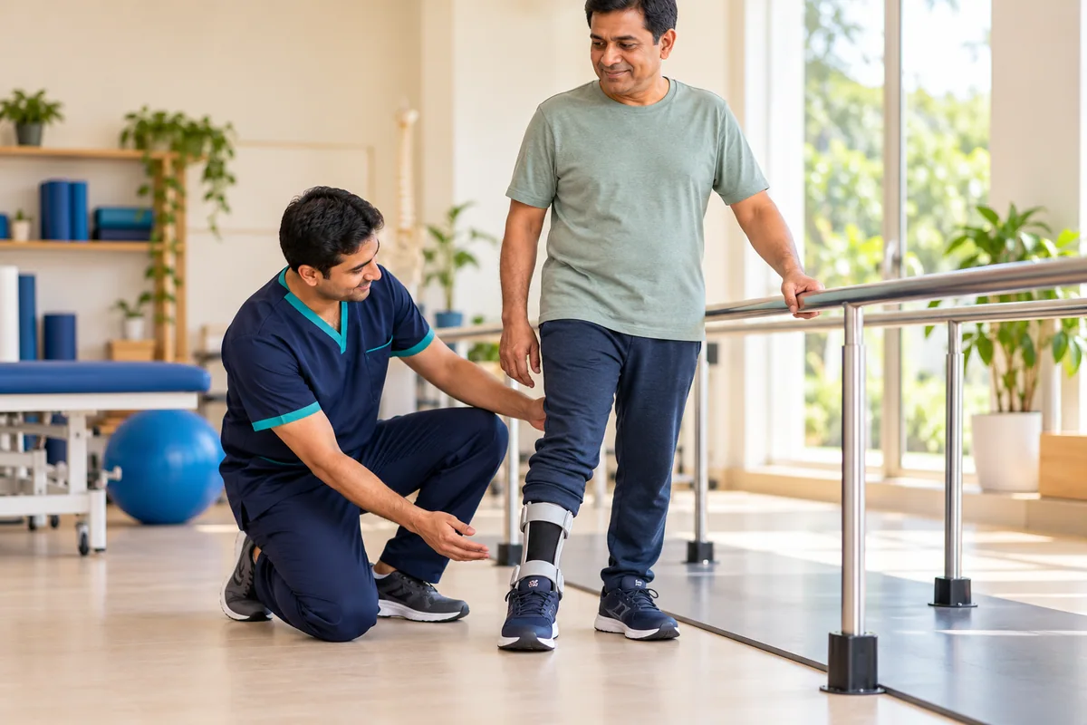
The featured image and supporting patient illustrations are AI-generated and do not depict actual patients.
ASPC Patient Education · Updated 26 October 2026
At a glance
Foot drop is difficulty lifting the front of the foot and toes. It is a sign of weakness rather than a diagnosis and may arise from a nerve, spinal or neurological condition. New weakness needs prompt medical assessment.
General patient information. Diagnosis and treatment should be individualized by a qualified healthcare professional.
Get help promptly: Sudden weakness with face, arm or speech changes: emergency care.
In this guide
- What Is Foot Drop?
- Causes and Risk Factors
- Symptoms and Functional Problems
- Clinical assessment and rehabilitation
- What the assessment includes
- How the care plan is chosen
- Recovery Time and Prognosis
- Nutrition and Daily Tips
- Other Conditions and When to Get Help
- Final Takeaway
- Common questions
- Sources and further reading
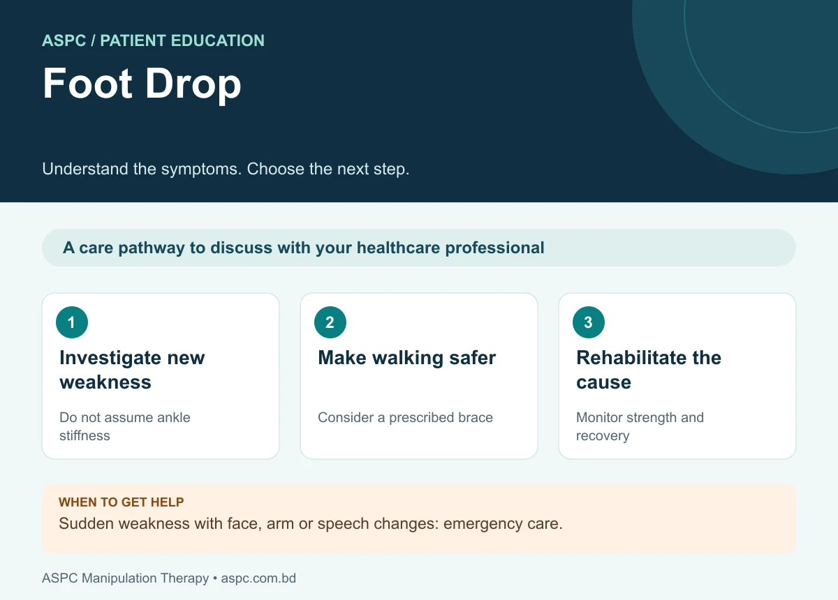
- Investigate new weakness: Do not assume ankle stiffness.
- Make walking safer: Consider a prescribed brace.
- Rehabilitate the cause: Monitor strength and recovery.
Foot drop, sometimes called drop foot, describes difficulty lifting the front part of the foot and toes because of weakness of the ankle and toe dorsiflexors. During walking, the toes may catch the ground and the foot may slap down after heel contact. It is a clinical sign rather than a single disease, and the cause may lie in a peripheral nerve, nerve root, plexus, spinal cord, brain, or less commonly the muscles themselves.
The most useful first step is to identify the site and cause of the weakness. Appropriate neurological and musculoskeletal assessment, fall prevention, gait training and, when indicated, an ankle-foot orthosis (AFO) can improve safety and function while the underlying condition is treated. Recovery varies widely according to the cause, severity, duration and degree of nerve injury.
What Is Foot Drop?
Normal walking requires the ankle dorsiflexors—especially tibialis anterior, extensor hallucis longus and extensor digitorum longus—to lift the foot during swing and control the foot as it lowers after heel strike. Weakness of these muscles can produce a high-stepping gait, toe dragging, foot slap and compensatory hip or knee flexion. The deep fibular (peroneal) nerve supplies most of these muscles, while the L4–L5 nerve roots, sciatic nerve and lumbosacral plexus may also be involved.
Foot drop should not be treated as a diagnosis by itself. A similar walking pattern can arise from common fibular neuropathy near the fibular head, L5 radiculopathy, sciatic neuropathy, generalized peripheral neuropathy, stroke, spinal cord disease or a structural injury. Correct localization matters because management differs across these causes.
Causes and Risk Factors
Foot drop results when the neural pathway or muscles responsible for dorsiflexion cannot generate adequate force. The cause may be peripheral or central, acute or chronic, and sometimes more than one lesion is present.
- Common fibular nerve compression: The nerve is particularly exposed near the fibular head/neck. Pressure from leg crossing, prolonged kneeling or squatting, tight casts, positioning during surgery, trauma or a mass can affect its function.
- L5 nerve-root irritation or injury: A lumbar disc herniation, foraminal narrowing or other spinal pathology may impair dorsiflexion. Weak ankle inversion, supplied mainly by the tibial nerve but with L5 contribution, can help distinguish L5 radiculopathy from an isolated common fibular neuropathy.
- Sciatic nerve or lumbosacral plexus injury: Trauma, pelvic or hip surgery, compression, or other lesions proximal to the common fibular nerve can weaken dorsiflexion and may also affect additional muscles or sensory areas.
- Central nervous system disorders: Stroke, spinal cord disease, multiple sclerosis and other neurological conditions may affect descending motor pathways and produce weakness, altered tone or associated neurological signs.
- Generalized neuropathy or neuromuscular disease: Diabetic neuropathy, inherited neuropathies such as Charcot–Marie–Tooth disease, and selected muscle disorders may cause unilateral or bilateral foot drop.
- Direct injury and postoperative causes: Knee dislocation, fibular fracture, penetrating injury, and complications of procedures near the hip, knee or spine may damage the nerve or related structures.
Not everyone with these risk factors develops foot drop. Sudden weakness, especially after trauma or with severe back pain, saddle numbness, bladder or bowel dysfunction, rapidly progressing symptoms, or other new neurological deficits requires urgent medical assessment.
Symptoms and Functional Problems
Symptoms depend on the site and severity of the lesion. Consider professional assessment if you notice the following:
- Difficulty lifting the front of the foot or extending the toes
- Toe catching, tripping, or needing to lift the knee unusually high while walking
- Foot slap at initial contact and reduced control while lowering the foot
- Weakness of ankle dorsiflexion, toe extension and sometimes eversion
- Numbness, tingling or altered sensation over the outer lower leg or top of the foot
- Difficulty walking on the heel, climbing stairs, or walking safely on uneven ground
Pain is not always present. Pain radiating from the back may suggest a nerve-root problem; tenderness or sensory change near the fibular head may point toward common fibular neuropathy. Sensory patterns overlap, however, so no single symptom or bedside test should be used in isolation.
Safety note: new foot drop should be assessed promptly. Seek emergency care if it occurs with new bladder or bowel dysfunction, saddle-area numbness, rapidly worsening weakness, major trauma, or signs suggestive of stroke such as facial droop, arm weakness or speech difficulty.
Clinical assessment and rehabilitation

Related background: plid and disc prolapse guide and diabetic neuropathy guide.
What the assessment includes
A systematic assessment considers contractile structures (muscle and tendon), non-contractile structures (joint and ligament), neural function, gait and functional safety. For foot drop, the neurological examination is essential because ankle stiffness or local tissue pain alone cannot explain all cases.
- History and onset: record when weakness began, whether it is progressing, trauma or surgery, back or leg pain, prolonged compression, weight loss, diabetes, medication and previous neurological disease.
- Gait and functional assessment: observe toe clearance, steppage gait, foot slap, balance, walking speed, stair negotiation, footwear and falls risk.
- Muscle testing and joint movement: assess ankle dorsiflexion, great-toe extension, toe extension, eversion, inversion and plantarflexion; measure ankle range and screen for contracture. Compare both sides.
- Neurological localization: test sensation and reflexes, and examine proximal muscles such as hip abductors. Inversion weakness can support L5 root involvement, while a pattern limited to fibular-innervated muscles may suggest common fibular neuropathy.
- Investigations and referral: a clinician may request nerve-conduction studies and electromyography (NCS/EMG), lumbar or peripheral nerve imaging, or other tests depending on the suspected cause. New or progressive weakness warrants prompt medical evaluation.
How the care plan is chosen
- Diagnosis-led plan: management follows the identified lesion, severity, irritability and functional goals; a single protocol is not suitable for every patient.
- Gait training and strengthening: when safe and appropriate, train controlled dorsiflexion, active-assisted movement, balance and task-specific walking. Strengthening must target muscles with preserved or recovering innervation; exercise cannot reverse every nerve lesion.
- Ankle-foot orthosis (AFO): an appropriately fitted brace can improve toe clearance, reduce tripping and stabilize the ankle during gait. Skin checks and reassessment are important, especially with sensory loss.
- Electrical stimulation: functional electrical stimulation may be considered for selected patients, particularly some central neurological conditions, after assessment of the nerve pathway and device suitability.
- Manual therapy and mobility: hands-on techniques may be used for associated joint stiffness, pain or soft-tissue restriction, but they do not by themselves repair a damaged nerve. Avoid forceful treatment where fracture, acute injury or serious neurological pathology is suspected.
- Regular reassessment and co-management: track dorsiflexion strength, sensation, gait, falls, skin condition and functional goals; coordinate with neurology, orthopaedics, spine services or rehabilitation medicine as indicated.
Recovery Time and Prognosis
There is no single recovery timeline. Prognosis depends on the cause, degree of nerve compression or axonal injury, duration before treatment, electrodiagnostic findings, and whether the underlying lesion can be corrected. Some compressive neuropathies improve after pressure is removed and rehabilitation begins, while severe nerve injury or central neurological disease may leave persistent weakness.
The practical goals are safe walking, adequate toe clearance, prevention of falls and contracture, maintenance of ankle range, and recovery of strength where possible. AFO use and compensatory gait strategies may remain useful even while nerve recovery is being monitored.
Nutrition and Daily Tips
No specific food cures foot drop. A balanced diet with adequate protein, vegetables, fruits, whole grains and fluids supports general health and rehabilitation. Where diabetes or another systemic condition contributes to neuropathy, individualized medical and nutritional management may be important.
Avoid prolonged pressure over the fibular head, including habitual leg crossing or sustained deep squatting when these provoke symptoms. Wear supportive footwear, keep walking areas clear, use prescribed walking aids, check skin beneath an orthosis, and follow the home exercise plan. Do not drive or undertake risky activities if foot control is insufficient for safe operation.
Other Conditions and When to Get Help
Not every case of toe dragging is caused by a common fibular nerve lesion. Differential diagnoses include L5 radiculopathy, sciatic neuropathy, lumbosacral plexopathy, stroke, spinal cord disease, generalized neuropathy, tendon injury, pain-limited movement and severe ankle restriction. Prompt assessment is needed to distinguish these conditions.
- New difficulty lifting the foot or toes, even if pain is absent
- Weakness that is worsening, spreading, or affecting both legs
- Foot drop after trauma, knee dislocation, fracture or recent surgery
- Severe back pain with saddle numbness, or new bladder/bowel dysfunction (emergency)
- Sudden weakness with facial droop, arm weakness, speech or vision changes (emergency)
- Repeated falls, skin injury from a brace, or inability to walk safely
Final Takeaway
Foot drop is a functional sign of impaired ankle and toe dorsiflexion, not a diagnosis in itself. The most important step is to identify where the motor pathway is affected—often the common fibular nerve or L5 nerve root, but sometimes a more proximal or central neurological structure. Accurate assessment, timely medical investigation, gait-focused physiotherapy, fall prevention and an appropriately fitted AFO can improve safety and function. Knowledge of the dorsiflexor muscle attachments and ankle ligament anatomy helps the clinician examine local structures without overlooking the neural cause. New or progressive foot drop requires prompt professional assessment.
Common questions
Is foot drop just a weak ankle?
No. It may result from a peripheral nerve problem, nerve-root compression, stroke or another disorder. Identifying the cause is essential.
Can a brace help?
An ankle-foot orthosis may improve toe clearance and walking safety in selected people. It should be fitted and reviewed by an appropriate professional.
Will every case fully recover?
Recovery depends on the underlying condition and extent of nerve or muscle damage. Some people need long-term support and rehabilitation.
Assessment and rehabilitation in Dhaka
ASPC Manipulation Therapy provides physiotherapy assessment and rehabilitation in Dhaka. An appointment can help clarify your symptoms, current movement and strength, daily activity goals and whether medical referral is needed. Care should be individualized, with progress reviewed rather than a guaranteed recovery promise.
ASPC describes its assessment framework as Structural Diagnosis and Management (SDM). A named framework does not establish effectiveness for every condition. Treatment choices should follow the clinical findings, relevant guidance and the person’s preferences.
Discuss an assessment with ASPC
Related patient guides
- Heel Pain: Causes, Plantar Fasciitis and Rehabilitation
- Ankle Sprain: Symptoms, Early Care and Rehabilitation
Sources and further reading
Read the condition-specific guidance below alongside the assessment and exercise information in this article. Guidelines inform general care; they do not establish individual outcomes or superiority of a clinic.
Editorial update: 26 October 2026. Medical information disclaimer.