

The featured image and supporting patient illustrations are AI-generated and do not depict actual patients.
ASPC Patient Education · Updated 26 October 2026
At a glance
An ankle sprain injures one or more ankle ligaments, usually after a twist. Rehabilitation progresses from protection and comfortable movement to strengthening, balance and a gradual return to daily activity or sport.
General patient information. Diagnosis and treatment should be individualized by a qualified healthcare professional.
Get help promptly: Deformity, a cold or numb foot or inability to bear weight needs prompt medical care.
In this guide
- What Is an Ankle Sprain?
- Causes and Risk Factors
- Symptoms and Pain Location
- Grades of Injury and Recovery Time
- Clinical assessment and rehabilitation
- What the assessment includes
- How the care plan is chosen
- Nutrition and Daily Tips
- Other Conditions and When to Get Help
- Final Takeaway
- Common questions
- Sources and further reading
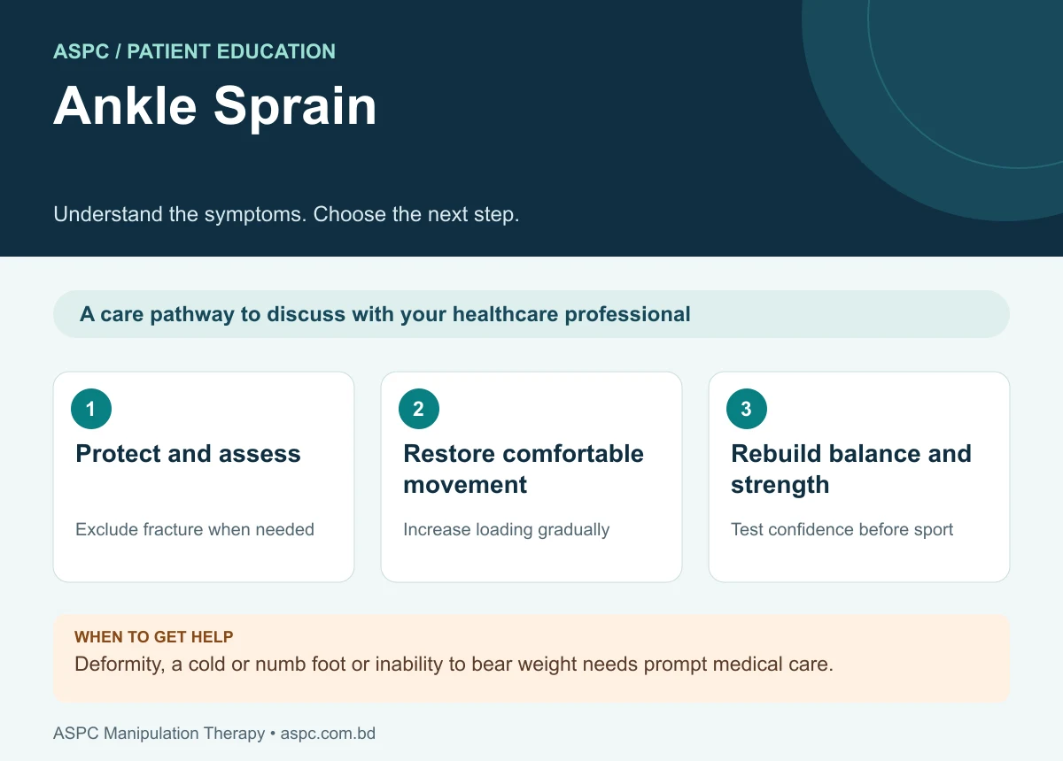
- Protect and assess: Exclude fracture when needed.
- Restore comfortable movement: Increase loading gradually.
- Rebuild balance and strength: Test confidence before sport.
An ankle sprain is an injury to one or more ligaments that support the ankle and foot, most commonly after the foot rolls inward during landing, walking on uneven ground or changing direction during sport. It can cause pain, swelling, bruising and difficulty bearing weight. The severity ranges from microscopic ligament damage to a complete tear.
Most people improve with appropriate protection, progressive loading and rehabilitation. However, recurrent sprains and a feeling that the ankle “gives way” can persist when strength, balance and movement control are not fully restored. Assessment should therefore address both the injured ligament and the function of the whole lower limb.
What Is an Ankle Sprain?
The ankle complex includes the talocrural joint, formed by the tibia, fibula and talus, and the subtalar joint between the talus and calcaneus. Ligaments provide passive stability, while muscles and tendons control movement and dynamic balance. A sprain occurs when ligament fibres are stretched or torn beyond their tolerance. Lateral ankle sprains are the most common and usually involve the anterior talofibular ligament (ATFL), with the calcaneofibular ligament (CFL) sometimes affected in more severe injuries.
The medial deltoid ligament resists excessive eversion, while the distal tibiofibular syndesmosis stabilizes the mortise between the tibia and fibula. Syndesmotic (“high ankle”) sprains usually follow external rotation or forced dorsiflexion and may take longer to recover than uncomplicated lateral sprains.
Causes and Risk Factors
- Inversion with plantarflexion: the common mechanism places tensile stress on the ATFL, and potentially the CFL if the force is greater.
- Sport and sudden direction changes: jumping, landing on another player’s foot, cutting and rapid acceleration can overload the ligament complex.
- Previous sprain: incomplete recovery can leave reduced dorsiflexion, strength, balance or confidence and increase recurrence risk.
- Muscle and sensorimotor deficits: reduced evertor strength, impaired proprioception and poor single-leg postural control can affect dynamic stability.
- External rotation / forced dorsiflexion: may injure the distal tibiofibular syndesmosis rather than the usual lateral ligament complex.
Not every ankle injury is a simple sprain. Fracture, tendon injury, osteochondral injury and syndesmotic injury may produce similar symptoms. Marked swelling, bony tenderness or inability to take weight should prompt a structured assessment and consideration of imaging.
Symptoms and Pain Location
Pain, swelling and reduced function are typical. Symptoms depend on the ligament involved and injury severity.
- Pain and tenderness: often around the outer ankle near the ATFL; medial pain may suggest deltoid injury, while pain above the ankle joint may suggest syndesmosis involvement.
- Swelling and bruising: may appear within hours and can spread toward the foot.
- Difficulty bearing weight: walking, stairs, running and single-leg standing may be painful or limited.
- Reduced movement or strength: ankle dorsiflexion, plantarflexion, inversion or eversion may be limited by pain and swelling.
- Instability: the ankle may feel unreliable or “give way,” particularly during turning or on uneven surfaces.
Urgent medical assessment is appropriate after major trauma, deformity, severe or rapidly increasing pain, numbness, a cold or discoloured foot, an open wound, or inability to take four steps. Persistent pain or swelling, locking, or repeated giving-way also requires reassessment.
Grades of Injury and Recovery Time
- Grade I (mild): ligament fibres are stretched or have small tears, with mild swelling and little functional loss. Many people improve over days to a few weeks.
- Grade II (moderate): partial tearing causes more swelling, bruising, pain and difficulty walking. Recovery commonly takes several weeks and should be guided by function.
- Grade III (severe): complete rupture or major instability causes substantial symptoms; recovery may take several weeks to months and may require specialist assessment.
These time ranges are approximate, not guarantees. Recovery depends on the exact structure injured, severity, previous sprains, general health, rehabilitation participation and the demands of work or sport. Persistent symptoms can develop into chronic ankle instability, so return to activity should be based on function rather than time alone.
Clinical assessment and rehabilitation

What the assessment includes
- History and mechanism: record the direction of injury, time since injury, ability to bear weight, previous sprains, sport/work demands and any red-flag symptoms.
- Structural examination: inspect swelling and bruising; palpate relevant ligaments and bones; measure active and passive range of motion, especially dorsiflexion; assess calf, evertor and invertor strength and gait.
- Ligament and functional testing: when appropriate and tolerable, use validated clinical tests such as the anterior drawer and talar tilt; assess balance and functional tasks progressively. Ottawa Ankle Rules can help determine whether radiographs are indicated after acute injury.
- Differential diagnosis: consider fracture, syndesmotic injury, tendon injury, osteochondral lesion and other causes of persistent ankle pain. Imaging is selected according to clinical findings and decision rules.
How the care plan is chosen
- Protection and optimal loading: use an appropriate brace or taping and crutches if needed, then progressively increase weight-bearing as symptoms allow. Severe sprains may need brief immobilization, generally no longer than about 10 days unless a clinician identifies another reason.
- Exercise-led rehabilitation: begin protected range-of-motion exercises and progress to calf/evertor strengthening, neuromuscular training, single-leg balance, hopping and task-specific drills. Exercise should be tailored to findings and healing stage.
- Manual therapy: joint and soft-tissue techniques may be used alongside exercise to help pain-free movement, swelling and dorsiflexion; they should not replace progressive loading and functional training.
- Regular reassessment: track pain, swelling, dorsiflexion, strength, balance, gait and patient-reported function, then adjust the program as recovery progresses.
- Return to sport/work: consider pain, ankle impairments, confidence, sensorimotor control and sport-specific performance, consistent with the PAASS framework.
Nutrition and Daily Tips
No particular food heals a sprained ligament on its own. A balanced diet with adequate protein, fruits, vegetables, whole grains and fluids supports general recovery. Avoid returning too quickly to running or jumping simply because pain has eased. Follow the prescribed loading plan, use support as advised, elevate the ankle when swollen, and perform home exercises consistently. Ice wrapped in a cloth may provide short-term symptom relief, but it should not delay assessment or rehabilitation.
Other Conditions and When to Get Help
An ankle sprain can resemble fracture, Achilles tendon injury, peroneal tendon injury, syndesmotic injury or an osteochondral lesion. Seek prompt medical assessment if you cannot take four steps after the injury, have bone tenderness at the malleoli or midfoot, notice deformity, develop numbness or a cold/discoloured foot, or have severe pain. Arrange reassessment if pain, swelling or instability is not improving as expected or if the ankle repeatedly gives way.
Final Takeaway
An ankle sprain is a ligament injury that can range from mild stretching to a severe tear. Understanding the ligament attachments and the dynamic roles of the surrounding muscles helps explain pain, instability and rehabilitation needs. Most cases benefit from appropriate protection, progressive weight-bearing and structured exercise, with balance and sport-specific training to reduce recurrence. Severe symptoms, inability to bear weight or persistent instability warrant professional assessment. The goal is not simply less pain, but a stable, strong and confident return to everyday activity and sport.
Common questions
Is every twisted ankle a sprain?
No. Fracture, tendon injury and a high ankle sprain can produce similar symptoms. Marked pain or inability to bear weight warrants assessment.
How long does an ankle sprain take to recover?
Recovery varies with severity and associated injury. Function, swelling, strength and balance are more useful guides than a fixed return date.
Why does the ankle keep giving way?
Persistent weakness, balance deficits or ligament instability may contribute. A structured rehabilitation programme and reassessment can help identify the problem.
Assessment and rehabilitation in Dhaka
ASPC Manipulation Therapy provides physiotherapy assessment and rehabilitation in Dhaka. An appointment can help clarify your symptoms, current movement and strength, daily activity goals and whether medical referral is needed. Care should be individualized, with progress reviewed rather than a guaranteed recovery promise.
ASPC describes its assessment framework as Structural Diagnosis and Management (SDM). A named framework does not establish effectiveness for every condition. Treatment choices should follow the clinical findings, relevant guidance and the person’s preferences.
Discuss an assessment with ASPC
Related patient guides
- Heel Pain: Causes, Plantar Fasciitis and Rehabilitation
- Foot Drop: Causes, Warning Signs and Rehabilitation
Sources and further reading
Read the condition-specific guidance below alongside the assessment and exercise information in this article. Guidelines inform general care; they do not establish individual outcomes or superiority of a clinic.
Editorial update: 26 October 2026. Medical information disclaimer.


