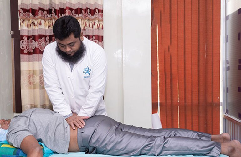

The featured image and supporting patient illustrations are AI-generated and do not depict actual patients.
ASPC Patient Education · Updated 26 October 2026
At a glance
Proximal neuropathy can cause pain and weakness around the hip and thigh, sometimes followed by muscle wasting. Diabetic amyotrophy is one form. Progressive weakness needs medical or neurological investigation.
General patient information. Diagnosis and treatment should be individualized by a qualified healthcare professional.
Get help promptly: Rapidly worsening weakness, saddle numbness or bladder changes need emergency assessment.
In this guide
- What Is Proximal Neuropathy?
- How Common Is It? Prevalence and Epidemiology
- Causes and Risk Factors
- Symptoms and Pain Location
- Stages and Recovery Time
- Assessment and Treatment: Structural Diagnosis and Management (SDM) at ASPC Manipulation Therapy
- Muscle and Ligament Attachments Around the Ankle
- Nutrition and Daily Tips
- Other Conditions and When to Get Help
- Proximal Neuropathy Assessment in Dhaka: Agrani Specialized Physiotherapy Centre (ASPC)
- Final Takeaway
- Common questions
- References and clinical guidance
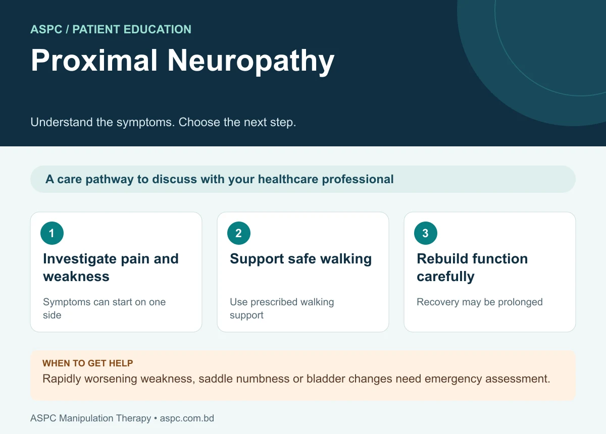
- Investigate pain and weakness: Symptoms can start on one side.
- Support safe walking: Use prescribed walking support.
- Rebuild function carefully: Recovery may be prolonged.
Proximal neuropathy is a pattern of peripheral nerve injury that causes pain, weakness and sometimes muscle wasting close to the trunk, most often around the hip and thigh when the lumbosacral region is involved. One important form is lumbosacral radiculoplexus neuropathy, often called diabetic amyotrophy when associated with diabetes. It can make standing, climbing stairs, rising from a chair or walking difficult (Dyck & Windebank, 2002).

The featured image and supporting patient illustrations are AI-generated and do not depict actual patients.
Symptoms often begin over days or weeks and may progress before gradually stabilizing. Because spinal, plexus, nerve, muscle and systemic conditions can produce similar symptoms, assessment should establish the distribution and cause rather than assume that all proximal pain in a person with diabetes is diabetic neuropathy (Glenn & Jabari, 2020).
What Is Proximal Neuropathy?
“Proximal neuropathy” is a descriptive term rather than one single diagnosis. In clinical practice it commonly refers to a painful, asymmetric neuropathy affecting nerve roots, the lumbosacral plexus and peripheral nerves supplying the hip and thigh. Diabetic lumbosacral radiculoplexus neuropathy (DLRPN), also known as diabetic amyotrophy or Bruns–Garland syndrome, is a recognized subtype; a similar syndrome can occur in people without diabetes (Dyck & Windebank, 2002; Thaisetthawatkul & Dyck, 2010).
The disorder is thought to involve axonal injury and an inflammatory microvasculitic process in affected nerves. It is different from the more common distal symmetric diabetic polyneuropathy, which typically begins with numbness or burning in the toes and feet on both sides. Proximal neuropathy is often markedly asymmetric at onset and may involve severe thigh or hip pain followed by weakness and wasting .
How Common Is It? Prevalence and Epidemiology
Lumbosacral radiculoplexus neuropathy is uncommon. A population-based study estimated an incidence of approximately 4.16 cases per 100,000 people per year; estimates vary by diagnostic criteria and study population. Diabetes is an important risk factor, but the condition can also occur without diabetes (Ng et al., 2019; Thaisetthawatkul & Dyck, 2010).
| Group / feature | Clinical interpretation |
|---|---|
| General population | Uncommon; population-based incidence about 4.16 per 100,000 person-years in one study |
| People with diabetes | Risk is substantially higher than in people without diabetes; diabetes is not required for diagnosis |
| Typical presentation | Middle-aged or older adults; often acute-to-subacute, painful and asymmetric |
| Course | Usually monophasic, with progression over weeks to months followed by gradual recovery; residual weakness may persist |
The condition does not simply reflect poor glucose control or long-standing diabetes. It may appear relatively near the time diabetes is diagnosed, and its mechanism differs from the chronic length-dependent nerve injury typical of distal symmetric diabetic neuropathy. Clinical history and examination remain central, with electrodiagnostic studies helping localize the process .
Causes and Risk Factors
Proximal neuropathy has several possible causes. The label should be refined after clinical assessment because compression, inflammatory disease, tumor, infection and other disorders can mimic a radiculoplexus neuropathy (Glenn & Jabari, 2020; Thaisetthawatkul & Dyck, 2010).
- Diabetes: diabetic lumbosacral radiculoplexus neuropathy is a well-recognized association, but it can occur even when diabetes has been diagnosed recently or glycemic control appears reasonable.
- Non-diabetic inflammatory radiculoplexus neuropathy: a clinically similar syndrome may occur without diabetes; both forms can cause pain, asymmetric weakness and muscle atrophy (Dyck & Windebank, 2002).
- Postsurgical inflammatory neuropathy: some neuropathies begin after surgery and may resemble a focal or multifocal nerve injury; timing and distribution guide investigation.
- Structural or infiltrative conditions: lumbar spinal disease, pelvic or retroperitoneal masses, malignancy, radiation injury, infection and systemic inflammatory disorders must be considered when the presentation is atypical or progressive.
- Weight loss and systemic symptoms: unintentional weight loss is commonly reported with DLRPN and should be documented, while also prompting consideration of alternative systemic causes.
Risk factors do not establish the diagnosis. New progressive weakness, marked asymmetry, severe pain or unexplained weight loss warrants timely medical assessment rather than treatment based on presumed muscle tightness alone.
Symptoms and Pain Location
Pain, weakness and loss of function are common features. Symptoms often begin on one side, around the hip, buttock or front of the thigh, and can spread over weeks or months (Glenn & Jabari, 2020).
- Deep aching, burning or lancinating pain: in the hip, buttock, groin or thigh, sometimes beginning before weakness.
- Progressive weakness: in hip flexion, hip adduction or knee extension, depending on the nerves involved.
- Muscle wasting: around the thigh or hip when denervation and reduced use persist.
- Reduced knee reflex: or other reflex changes, depending on the distribution of nerve involvement.
- Sensory changes: such as altered sensation or tingling; sensory symptoms may be less prominent than pain and weakness.
- Walking and transfer difficulty: including climbing stairs, standing from a chair, maintaining balance or walking without support.
- Spread of symptoms: to the other leg or more distal muscles; foot drop can occur in some patients.
Proximal neuropathy can affect the ankle and foot indirectly if more distal nerve fibers are involved. Foot drop, for example, may occur when weakness extends to muscles that lift the foot. However, isolated ankle pain or ligament tenderness is not enough to diagnose proximal neuropathy and may indicate a separate musculoskeletal condition .
Urgent warning: seek emergency assessment for sudden severe weakness, new loss of bladder or bowel control, numbness around the saddle region, rapidly progressive bilateral symptoms, or severe back pain with fever or major trauma. These can indicate conditions requiring immediate investigation.
Stages and Recovery Time
Proximal neuropathy has no universally accepted three named stages. Its course is more usefully described as an acute or subacute painful phase, evolving weakness, and gradual recovery. These phases overlap and vary between people (Dyck & Windebank, 2002; Glenn & Jabari, 2020).
- Early painful phase: hip, buttock or thigh pain develops over days to weeks; pain may be severe and may precede obvious weakness.
- Progressive weakness phase: weakness, reduced reflexes and muscle wasting become more apparent. Symptoms can spread to additional nerve territories or the opposite leg over weeks to months.
- Recovery phase: pain often improves before strength and muscle bulk recover. Improvement may continue for many months, and some people retain weakness, sensory symptoms or gait limitations.
There is no single recovery timeline. The extent of axonal injury, severity of weakness, other health conditions, falls, deconditioning and the presence of alternative or coexisting diagnoses all influence recovery. Many patients improve, but recovery can be prolonged and incomplete; rehabilitation should therefore prioritize safety, mobility and gradual restoration of function (Thaisetthawatkul & Dyck, 2010; Glenn & Jabari, 2020).
Assessment and Treatment: Structural Diagnosis and Management (SDM) at ASPC Manipulation Therapy
Treatment should begin with an accurate diagnosis and a safety-focused neurological assessment. At ASPC Manipulation Therapy in Dhaka, the published Structural Diagnosis and Management (SDM) framework describes systematic assessment of musculoskeletal and neurological function. For proximal neuropathy, such an approach can organize examination, but it does not replace medical or neurological investigation when a nerve disorder is suspected (Haque et al., 2020; Glenn & Jabari, 2020).
How SDM assesses proximal weakness and pain
A structured assessment should combine history, neurological examination and functional evaluation. Because radiculoplexus neuropathy affects nerve roots, plexus and peripheral nerve territories, assessment must not be limited to joint movement or muscle length alone .
- History and symptom mapping: record onset, pain distribution, weakness progression, weight change, diabetes status, recent surgery, cancer history, infection symptoms and functional changes.
- Neurological examination: test key muscle groups, reflexes, sensation and gait; compare sides and document whether weakness follows a single root, a named nerve or a multifocal pattern.
- Structural and functional examination: assess hip, knee, ankle and foot movement, muscle length, balance, transfers and walking, while distinguishing pain-limited movement from true neurological weakness.
- Investigations and differential diagnosis: nerve-conduction studies and needle electromyography can help localize axonal injury. MRI of the lumbar spine or plexus and laboratory testing may be indicated to exclude compression, malignancy, infection or systemic disease.
How SDM guides management
- Diagnosis-led plan: the program should be based on the confirmed or most likely cause, severity of weakness, pain irritability and current functional capacity.
- Medical management: pain control and treatment of diabetes or another underlying disorder should be coordinated with the treating physician or neurologist. Immunotherapy has been considered in selected cases, but evidence is limited and treatment decisions require specialist judgment.
- Physiotherapy and rehabilitation: use graded strengthening when tolerated, range-of-motion exercises, gait and balance training, transfer practice and conditioning. Avoid aggressive loading of profoundly weak muscles or techniques that worsen pain.
- Assistive devices and fall prevention: a cane, walker, ankle-foot orthosis for foot drop, home-safety modifications or supervised mobility may be needed temporarily.
- Regular reassessment: repeat strength, reflex, sensation, pain, walking and daily-function measures; adjust the plan as symptoms evolve.
Evidence context: SDM has a published description concerning lumbar mechanical dysfunction and a journal-published trial concerning plantar heel pain. These are different study designs and clinical populations. They do not establish that SDM is superior or effective for every presentation of proximal neuropathy. Care should follow the diagnosis, relevant guidance and measured progress; medical referral takes priority when indicated.
Muscle and Ligament Attachments Around the Ankle
Although proximal neuropathy primarily affects the hip and thigh, neurological involvement may extend distally and alter ankle control. The following concise anatomy reference is included to support examination of foot drop, ankle stability and gait. These attachments describe normal anatomy; they are not evidence that ankle ligaments are the cause of proximal neuropathy .
| Structure | Origin / proximal attachment | Insertion / distal attachment | Main action or role |
|---|---|---|---|
| Tibialis anterior (muscle) | Lateral condyle and upper lateral tibia; interosseous membrane | Medial cuneiform and base of 1st metatarsal | Dorsiflexion and inversion |
| Extensor hallucis longus (muscle) | Middle anterior fibula and interosseous membrane | Base of distal phalanx of great toe | Extends great toe; assists dorsiflexion |
| Extensor digitorum longus (muscle) | Lateral tibial condyle, anterior fibula and interosseous membrane | Middle and distal phalanges of toes 2–5 via extensor expansions | Extends toes; dorsiflexes ankle |
| Fibularis (peroneus) longus | Head and upper lateral fibula | Plantar surface of medial cuneiform and base of 1st metatarsal | Eversion; assists plantarflexion and supports transverse arch |
| Fibularis brevis | Lower lateral fibula | Tuberosity at base of 5th metatarsal | Eversion; assists plantarflexion |
| Gastrocnemius and soleus (triceps surae) | Femoral condyles (gastrocnemius); posterior proximal tibia/fibula (soleus) | Calcaneus via calcaneal/Achilles tendon | Plantarflexion; gastrocnemius also flexes knee |
| Tibialis posterior | Posterior tibia and fibula; interosseous membrane | Navicular tuberosity and multiple midfoot bones | Inversion, plantarflexion and medial arch support |
| Anterior talofibular ligament (ATFL) | Anterior margin of lateral malleolus (fibula) | Neck of talus | Resists anterior talar translation, especially in plantarflexion |
| Calcaneofibular ligament (CFL) | Tip of lateral malleolus | Lateral calcaneus | Resists inversion; supports ankle and subtalar stability |
| Posterior talofibular ligament (PTFL) | Malleolar fossa of lateral malleolus | Posterior talus | Contributes to lateral ankle stability |
| Deltoid ligament complex | Medial malleolus | Talus, calcaneus and navicular via multiple bands | Resists eversion and stabilizes medial ankle |
| Anterior inferior tibiofibular ligament (AITFL) | Anterolateral distal tibia | Anterior distal fibula | Stabilizes distal tibiofibular syndesmosis |
Clinical relevance: the deep fibular nerve supplies the principal ankle dorsiflexors, while the superficial fibular nerve supplies the fibularis muscles. Weak dorsiflexion may cause foot drop and toe catching; eversion weakness can further reduce lateral ankle control. Ligaments provide passive stability, whereas muscles and tendons provide active control. Examination should assess both systems and investigate the neurological cause when weakness is present .
Nutrition and Daily Tips
No specific food cures proximal neuropathy. A balanced diet with adequate protein, vegetables, fruit, whole grains and fluids supports general health and rehabilitation. Unintentional weight loss should be reported to a clinician rather than managed only with dietary changes. If diabetes is present, individualized nutritional and glucose-management advice should be coordinated with the healthcare team .
Use prescribed walking aids, clear trip hazards, wear stable footwear, inspect the feet if sensation is reduced, and pace activity to avoid falls and excessive fatigue. Do not begin intense strengthening or manipulation without assessment when weakness is progressive or the diagnosis is uncertain.
Other Conditions and When to Get Help
Not all hip or thigh pain with weakness is proximal neuropathy. Lumbar radiculopathy, femoral neuropathy, hip joint disease, spinal stenosis, myopathy, motor neuron disease, malignancy and other plexopathies may resemble it. Seek prompt medical assessment if pain is severe or persistent, weakness is increasing, walking becomes unsafe, symptoms spread, or unexplained weight loss occurs (Glenn & Jabari, 2020).
- Rapidly progressive weakness or inability to walk safely: arrange urgent or prompt clinical review according to severity.
- New bladder or bowel dysfunction or saddle numbness: arrange urgent or prompt clinical review according to severity.
- Fever, recent serious infection, cancer history or major unexplained weight loss: arrange urgent or prompt clinical review according to severity.
- New foot drop, repeated falls or significant loss of sensation: arrange urgent or prompt clinical review according to severity.
- Symptoms following surgery or trauma, or symptoms that do not fit a typical pattern: arrange urgent or prompt clinical review according to severity.
Sudden severe symptoms, bladder/bowel changes, saddle anesthesia or rapidly worsening bilateral weakness require emergency assessment.
Proximal Neuropathy Assessment in Dhaka: Agrani Specialized Physiotherapy Centre (ASPC)
Final Takeaway
Proximal neuropathy is a potentially disabling nerve disorder characterized by pain and asymmetric weakness around the hip and thigh, sometimes extending to the lower leg and ankle. Diabetic lumbosacral radiculoplexus neuropathy is an important form, but non-diabetic and other serious causes must also be considered. Diagnosis depends on careful history and neurological examination, often supported by electrodiagnostic testing and imaging. Treatment combines appropriate medical management with safe, progressive rehabilitation, gait support and fall prevention. Ankle muscle and ligament anatomy can help explain movement and stability findings, but it does not replace investigation of the underlying nerve disorder. Seek timely professional assessment for progressive pain, weakness or walking difficulty (Glenn & Jabari, 2020).
Common questions
Is proximal neuropathy the same as ordinary sciatica?
No. Symptoms may overlap, but the nerve distribution and underlying mechanism can differ. Neurological examination and sometimes nerve tests help distinguish them.
Can it occur without diabetes?
A similar radiculoplexus neuropathy can occur without diabetes. Other spinal, nerve and systemic causes also need consideration.
Can rehabilitation help?
Rehabilitation can support mobility, strength and fall prevention alongside medical treatment. It does not replace investigation or guarantee full nerve recovery.
Assessment and rehabilitation in Dhaka
ASPC Manipulation Therapy provides physiotherapy assessment and rehabilitation in Dhaka. An appointment can help clarify your symptoms, current movement and strength, daily activity goals and whether medical referral is needed. Care should be individualized, with progress reviewed rather than a guaranteed recovery promise.
ASPC describes its assessment framework as Structural Diagnosis and Management (SDM). A named framework does not establish effectiveness for every condition. Treatment choices should follow the clinical findings, relevant guidance and the person’s preferences.
Discuss an assessment with ASPC
Related patient guides
- Foot Drop: Causes, Warning Signs and Rehabilitation
- Diabetic Neuropathy: Symptoms, Foot Care and Rehabilitation
Related background: diabetic neuropathy guide.
Related assessment and rehabilitation guides
References and clinical guidance
Guidelines inform general care; studies have specific populations and limitations. The SDM framework description and plantar-heel-pain trial should be read in their own clinical context.
- NIDDK condition-specific patient guidance
- McCray, Stino, Davalos et al. (2025). Atypical diabetic neuropathies. BMJ.
- Dyck, Windebank (2002). Diabetic and nondiabetic lumbosacral radiculoplexus neuropathies: New insights into pathophysiology and treatment. Muscle & Nerve.
- Glenn, Jabari (2020). Diabetic Lumbosacral Radiculoplexus Neuropathy (Diabetic Amyotrophy). Neurologic Clinics.
- Haque (2020). Treating Mechanical Dysfunctions of Lower Back through Lumbar Spine Assessment of Structural Diagnosis and Management (SDM): A New Comprehensive Approach of Musculoskeletal Medicine. American Journal of Biomedical Science & Research.
- Ng, Dyck, Laughlin et al. (2019). Lumbosacral radiculoplexus neuropathy. Neurology.
- Thaisetthawatkul, Dyck (2010). Treatment of Diabetic and Nondiabetic Lumbosacral Radiculoplexus Neuropathy. Current Treatment Options in Neurology.
- Akter, Hossain, Hossain et al. (2024). Comparison of Structural Diagnosis and Management (SDM) approach and MyoFascial Release (MFR) for improving plantar heel pain, ankle range of motion and disability: A randomized clinical trial. Journal of Manual & Manipulative Therapy.
- Golanó et al. (2010): Anatomy of the ankle ligaments — pictorial essay
- Ng et al. (2019): population-based radiculoplexus neuropathy incidence study
Content restored and references checked: 26 October 2026. This editorial check does not replace individual clinical assessment. Medical information disclaimer.