

The featured image and supporting patient illustrations are AI-generated and do not depict actual patients.
ASPC Patient Education · Updated 26 October 2026
At a glance
A cervical rib is an extra rib near the base of the neck. Many cause no symptoms. In some people it contributes to compression of nerves or blood vessels, so treatment depends on the clinical findings.
General patient information. Diagnosis and treatment should be individualized by a qualified healthcare professional.
Get help promptly: A suddenly swollen, cold, pale or blue arm needs urgent medical assessment.
In this guide
- What Is a Cervical Rib?
- How Common Is It? Prevalence and Epidemiology
- Table 1. Pooled prevalence estimates reported by Henry et al. (2018).
- Causes and Risk Factors
- Muscle and Ligament Attachments Around the Cervical Rib
- Table 2. Key muscle and fibrous/ligamentous relationships relevant to cervical-rib assessment.
- Symptoms and Pain Location
- Diagnosis and Assessment
- Treatment and Physiotherapy Management
- Structural Assessment and Management (SDM)-Informed Approach
- Daily Tips and Self-Management
- Other Conditions and When to Get Help
- Cervical Rib Care: Choosing an Appropriate Service
- Final Takeaway
- Common questions
- References and clinical guidance
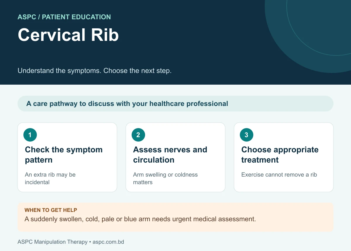
- Check the symptom pattern: An extra rib may be incidental.
- Assess nerves and circulation: Arm swelling or coldness matters.
- Choose appropriate treatment: Exercise cannot remove a rib.
A cervical rib is an additional rib-like bone that most often develops from the seventh cervical vertebra (C7). It is a congenital anatomical variant and is usually discovered incidentally. In a minority of people, it can contribute to pain, tingling, weakness or vascular symptoms by narrowing the thoracic outlet and compressing the brachial plexus or subclavian vessels (Henry et al., 2018).

The featured image and supporting patient illustrations are AI-generated and do not depict actual patients.
The presence of a cervical rib does not automatically mean that a person has a disorder or needs treatment. Clinical importance depends on the rib’s size and shape, associated fibrous bands, the relationship to nearby nerves and blood vessels, and the patient’s symptoms. Accurate assessment is therefore more useful than treating the radiographic finding alone (Henry et al., 2018).
What Is a Cervical Rib?
The cervical rib is a supernumerary rib arising most commonly from C7. It may be a short bony projection, an incomplete rib with a free tip, an incomplete rib connected to the first rib by a fibrous band, or a complete rib that articulates or fuses with the first rib. It can occur on one or both sides and varies considerably in length and shape .
A cervical rib lies close to the scalene muscles, brachial plexus and subclavian vessels. These structures pass through the thoracic outlet, a confined anatomical region between the neck and upper chest. A cervical rib or associated fibrous band can alter this space and may cause neurogenic or vascular thoracic outlet syndrome (TOS). Most cervical ribs, however, remain asymptomatic throughout life (Henry et al., 2018).
How Common Is It? Prevalence and Epidemiology
Reported prevalence varies according to the population studied and the imaging method used. A large meta-analysis of 141 studies involving 77,924 participants estimated cervical ribs in approximately 1.1% of healthy populations, compared with 29.5% among people assessed for thoracic outlet syndrome. These figures describe pooled study groups and should not be interpreted as the percentage of all people with cervical ribs who will develop symptoms (Henry et al., 2018).
| Group | Pooled cervical-rib prevalence |
|---|---|
| General/healthy populations | About 1.1% |
| People assessed for thoracic outlet syndrome | About 29.5% |
Table 1. Pooled prevalence estimates reported by Henry et al. (2018).
Cervical ribs are reported more often in women and are frequently unilateral, although either side or both sides may be affected. Differences between studies reflect variation in imaging, participant selection and definitions of a cervical rib. The rib is a risk factor for thoracic outlet compression, not proof that compression is present (Henry et al., 2018).
Causes and Risk Factors
A cervical rib is congenital and develops during embryological patterning of the vertebral column. Abnormal persistence or development of a cervical costal element, commonly at C7, produces the additional rib. It is not caused by poor posture, exercise or an injury acquired in adulthood, although trauma or repetitive activity may bring symptoms to attention in a person with this anatomical variant .
- Anatomical size and shape: a long or complete rib may create a more substantial bony boundary near the neurovascular structures, although symptom severity cannot be predicted from size alone.
- Fibrous bands: an associated fibrous connection between the cervical rib and first rib may tether or compress the lower brachial plexus or nearby vessels.
- Scalene muscle relationships: the anterior and middle scalene muscles form the sides of the interscalene triangle. Muscle anatomy and the insertion pattern can contribute to narrowing of this passage .
- Repetitive overhead activity or trauma: repeated arm elevation, heavy carrying or injury may aggravate symptoms in susceptible people, but these factors do not create the congenital rib itself .
Many people with a cervical rib never develop symptoms. Risk assessment should therefore focus on relevant clinical signs, functional limitation and objective evidence of nerve or vascular involvement rather than the image alone (Henry et al., 2018).
Muscle and Ligament Attachments Around the Cervical Rib
A cervical rib is an accessory bone, so its attachments are variable; it does not have one standard set of muscle and ligament attachments. In many cases, a fibrous band links the rib to the first rib near the anterior scalene insertion. The following structures are clinically important because of their close anatomical relationship to the cervical rib and thoracic outlet .
| Structure | Typical attachment / relationship | Clinical relevance |
|---|---|---|
| Anterior scalene | Cervical transverse processes, commonly C3–C6, to the scalene tubercle on the first rib; its insertion is near the potential fibrous connection of a cervical rib. | May form the anterior boundary of the interscalene triangle and influence neurovascular space. |
| Middle scalene | Cervical transverse processes, commonly C2–C7, to the superior surface of the first rib, posterior to the subclavian artery groove. | Forms the posterior boundary of the interscalene triangle. |
| Scalenus minimus (when present) | May extend from the lower cervical region, often C7, toward the first rib or suprapleural membrane. | An anatomical variant that can contribute to local crowding. |
| Fibrous band / anomalous ligament | May connect the cervical rib’s distal end to the first rib or nearby structures; configuration varies. | Can tether or compress the lower brachial plexus or subclavian vessels. |
| First rib and costoclavicular ligament region | The first rib is the inferior boundary of the interscalene triangle; the costoclavicular ligament connects the first rib to the clavicle. | Adjacent structures may influence thoracic-outlet dimensions; the ligament is not a universal direct attachment of the cervical rib. |
Table 2. Key muscle and fibrous/ligamentous relationships relevant to cervical-rib assessment.
Clinical anatomy note: the attachments above describe typical nearby anatomy and possible anomalous bands; they should not be read as fixed attachments present in every patient. If “ankle structure” was intended literally, ankle muscle and ligament attachments are anatomically unrelated to a cervical rib and should be covered in a separate document.
Symptoms and Pain Location
Most cervical ribs are asymptomatic. Symptoms occur when nearby nerves or blood vessels are compressed or irritated. Consider professional assessment if you notice the following :
- Pain or aching around the lower neck, shoulder, upper chest or arm
- Tingling or numbness, particularly along the inner forearm and ring and little fingers
- Hand weakness, reduced grip strength, clumsiness or difficulty with fine motor tasks
- Symptoms that worsen during prolonged overhead activity or certain arm positions
- Arm swelling, heaviness, bluish discoloration or visible venous distension
- Coldness, pallor, color change, reduced pulse or exertional pain in the hand
Neurological symptoms often involve the lower brachial plexus, particularly C8–T1-related function. Arterial compression may cause a cool or pale hand, reduced pulse or, in severe cases, digital ischemia. Venous obstruction can cause swelling and bluish discoloration. These patterns require assessment because other neurological and vascular disorders can produce similar symptoms .
Urgent warning: sudden arm swelling or discoloration, a cold or pale hand, severe pain, new marked weakness, or signs of impaired circulation require urgent medical evaluation. Do not assume these symptoms are harmless simply because a cervical rib is already known.
Diagnosis and Assessment
Assessment combines a detailed history, physical examination and imaging or physiological tests selected according to the suspected type of compression. The aim is to establish whether symptoms arise from a cervical rib and to exclude other causes, including cervical radiculopathy, peripheral nerve entrapment and shoulder disorders .
- History and functional review: ask about symptom location, duration, overhead activity, trauma, hand weakness, sensory changes, swelling, color change and effects on work, sleep and daily tasks.
- Neurological examination: assess sensation, myotomal strength, hand intrinsic muscle function, reflexes and possible lower brachial plexus involvement.
- Vascular examination: compare arm color, temperature, swelling, capillary refill and pulses. A normal pulse at rest does not fully exclude positional vascular compression.
- Provocative manoeuvres: positions such as elevated-arm or Adson-type tests may reproduce symptoms, but these tests have limited specificity and should not establish the diagnosis on their own .
- Imaging: plain radiographs can identify a cervical rib. CT can define bony anatomy, while MRI may assess soft tissues and the brachial plexus. Duplex ultrasound or vascular imaging may be indicated if vascular compression is suspected.
- Electrodiagnostic testing: nerve conduction studies and electromyography may help evaluate neurological dysfunction or distinguish it from other nerve disorders.
A cervical rib on an X-ray is not sufficient by itself to diagnose thoracic outlet syndrome. The imaging finding must be interpreted alongside symptoms, examination and, when needed, objective neurological or vascular investigations (Henry et al., 2018).
Treatment and Physiotherapy Management
Treatment depends on whether the cervical rib is asymptomatic, causes neurogenic symptoms or produces vascular compromise. An incidental rib without symptoms generally does not require treatment. For symptomatic patients without progressive neurological loss or significant vascular compromise, an initial conservative program may be appropriate, with specialist review when symptoms are persistent or concerning .
- Education and activity modification: temporarily reduce activities that reliably reproduce symptoms, particularly sustained overhead work, heavy shoulder loading or carrying loads that depress the shoulder girdle. Avoid unnecessary immobilization.
- Postural and movement retraining: address comfortable cervical and thoracic positioning, scapular control and graded tolerance for arm elevation without provoking neurological or vascular symptoms.
- Exercise: a physiotherapist may prescribe individualized mobility, breathing, scapular-control and low-load strengthening exercises. Exercises should be modified or stopped if they increase numbness, weakness, swelling, color change or coldness.
- Manual therapy: gentle techniques may be considered only after careful screening. Forceful cervical or first-rib manipulation should be avoided when vascular compromise, significant neurological deficit or other contraindications are suspected.
- Regular reassessment: track pain, sensation, strength, arm function and vascular signs. Worsening objective deficits or failure to improve should prompt medical reassessment rather than simply increasing exercise intensity.
- Medical and surgical management: analgesia or other medication is determined by a qualified prescriber. Surgery to remove the cervical rib and/or fibrous band may be considered for persistent, well-documented neurogenic compression despite conservative care, or for significant arterial or venous complications. Vascular involvement may require specialized vascular procedures .
The evidence for physiotherapy in thoracic outlet syndrome is variable, and management should be individualized. There is no single exercise program suitable for every cervical rib. Patients with signs of arterial or venous compromise need prompt specialist assessment rather than an exercise-only approach .
Structural Assessment and Management (SDM)-Informed Approach
A structural assessment framework can help organize examination of the cervical spine, shoulder girdle, muscle function and neurological status. In a patient with a cervical rib, it should be used as a clinical reasoning structure rather than as proof that a particular treatment method is effective. Published descriptions of the Structural Diagnosis and Management (SDM) approach do not establish cervical-rib-specific effectiveness; no cervical-rib outcome trial is cited here (Haque et al., 2020).
How a structured assessment can guide care
- History and red-flag screening: identify neurological and vascular symptoms, functional triggers, trauma history and signs that require urgent referral.
- Structural examination: compare cervical and shoulder movement, scapular mechanics, strength, sensation and symptom response without provoking sustained compression.
- Differential diagnosis: consider cervical radiculopathy, ulnar neuropathy, brachial plexus disorders, shoulder pathology and vascular disease.
How management is adjusted
- Diagnosis-led plan: choose exercise and activity modification according to the confirmed clinical pattern, irritability and functional goals.
- Avoid aggressive manipulation: a suspected cervical-rib-related neurovascular problem requires caution; treatment must not compromise blood flow or nerve function.
- Co-management and referral: coordinate with a physician, neurologist or vascular specialist when symptoms are progressive, objective deficits are present, or imaging suggests vascular involvement.
- Outcome monitoring: repeat functional, neurological and vascular checks and refer for further investigation if symptoms worsen or do not follow the expected course.
Daily Tips and Self-Management
No diet or supplement removes a cervical rib. General health habits may support rehabilitation, but the key steps are symptom-aware activity modification and appropriate clinical follow-up.
- Break up prolonged overhead tasks and keep frequently used items within comfortable reach.
- Avoid carrying heavy bags on a shoulder if this reliably worsens symptoms.
- Follow a prescribed exercise plan and do not push through tingling, weakness, swelling or color change.
- Maintain general physical activity within a symptom-safe range and arrange reassessment if function declines.
- Do not self-diagnose vascular compression from pulse changes alone or attempt forceful neck manipulation.
Other Conditions and When to Get Help
Symptoms associated with a cervical rib can resemble cervical disc disease, cervical radiculopathy, ulnar neuropathy, shoulder disorders and other causes of thoracic outlet syndrome. Seek medical assessment if symptoms persist, worsen, disturb sleep or affect hand function. Urgent evaluation is required for a cold or pale hand, sudden swelling or blue discoloration, severe pain, rapidly progressive weakness or other evidence of compromised circulation .
Cervical Rib Care: Choosing an Appropriate Service
Final Takeaway
A cervical rib is a congenital anatomical variant, usually arising from C7, and most people with one have no symptoms. In some cases, it can contribute to thoracic outlet compression affecting the brachial plexus or subclavian vessels. Diagnosis requires correlation of symptoms with examination and appropriate investigations, not an X-ray finding alone. Management ranges from reassurance and tailored physiotherapy to specialist or surgical treatment when significant neurological or vascular compromise is present. Early recognition of progressive weakness, a cold or discolored hand, or sudden swelling is important for safe care (Henry et al., 2018).
Common questions
Does every cervical rib need treatment?
No. An incidental rib without symptoms may need no active treatment. Symptoms require assessment to establish whether the rib is relevant.
Can physiotherapy remove a cervical rib?
No. Physiotherapy may help selected movement or nerve-related symptoms, but it does not remove the extra bone.
Is cervical rib the same as cervical spondylosis?
No. A cervical rib is a congenital extra rib; cervical spondylosis describes age-related changes in the neck. They should not be treated as interchangeable diagnoses.
Assessment and rehabilitation in Dhaka
ASPC Manipulation Therapy provides physiotherapy assessment and rehabilitation in Dhaka. An appointment can help clarify your symptoms, current movement and strength, daily activity goals and whether medical referral is needed. Care should be individualized, with progress reviewed rather than a guaranteed recovery promise.
ASPC describes its assessment framework as Structural Diagnosis and Management (SDM). A named framework does not establish effectiveness for every condition. Treatment choices should follow the clinical findings, relevant guidance and the person’s preferences.
Discuss an assessment with ASPC
Related patient guides
- Frozen Shoulder: Symptoms, Causes, Recovery and Treatment
- Neck Pain: Causes, Symptoms, Exercises and Physiotherapy
- Deltoid Tendinitis: Outer Shoulder Pain and Rehabilitation
Related background: neck pain guide.
References and clinical guidance
Guidelines inform general care; studies have specific populations and limitations. The SDM framework description and plantar-heel-pain trial should be read in their own clinical context.
- NHS condition-specific patient guidance
- Haque (2020). Treating Mechanical Dysfunctions of Lower Back through Lumbar Spine Assessment of Structural Diagnosis and Management (SDM): A New Comprehensive Approach of Musculoskeletal Medicine. American Journal of Biomedical Science & Research.
- Akter, Hossain, Hossain et al. (2024). Comparison of Structural Diagnosis and Management (SDM) approach and MyoFascial Release (MFR) for improving plantar heel pain, ankle range of motion and disability: A randomized clinical trial. Journal of Manual & Manipulative Therapy.
- Henry et al. (2018): cervical rib prevalence meta-analysis
Content restored and references checked: 26 October 2026. This editorial check does not replace individual clinical assessment. Medical information disclaimer.